How Are Nerve Impulses Transmitted
Each nerve has many extensions of individual nerve cells. These long, thread-like pieces are where nerve impulses are transmitted. A type of nerve cell that has a specific function to deliver messages to the brain is called a neuron. These messages are nerve impulses, and each message is a quick, electrical impulse. Neuron Structure. Nerve impulses have a domino effect. Each neuron receives an impulse and must pass it on to the next neuron and make sure the correct impulse continues on its path. Through a chain of chemical events, the dendrites (part of a neuron) pick up an impulse that’s shuttled through the axon and transmitted to the.
- How Are Nerve Impulses Transmitted Within A Neuron
- The Nerve Impulse
- How Are Nerve Impulses Transmitted From One Neuron To Another
- How Are Nerve Impulses Transmitted From One Neuron To Another
- How Are Nerve Impulses Transmitted From One Neuron To Another
Nerve Impulses
[Back to Nervous System]
| Resting Membrane Potential | Action Potentials | How impulses start (receptors) | Propagation of Impulses | Speed of Impulses |
Neurones send messages electrochemically; this means that chemicals (ions) cause an electrical impulse. Neurones and muscle cells are electrically excitable cells, which means that they can transmit electrical nerve impulses. These impulses are due to events in the cell membrane, so to understand the nerve impulse we need to revise some properties of cell membranes.

The Resting Membrane Potential[back to top]
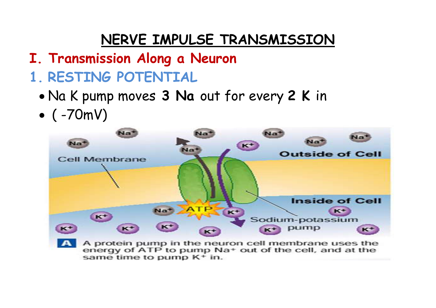
When a neurone is not sending a signal, it is at ‘rest’.The membrane is responsible for the different events that occur in a neurone.All animal cell membranes contain a protein pump called the sodium-potassium pump(Na+K+ATPase). This uses the energy from ATP splitting to simultaneously pump 3 sodium ions out of the cell and 2 potassium ions in.
The Sodium-Potassium Pump (Na+K+ATPase) Three sodium ions from inside the cell first bind to the transport protein. Then a phosphate group is transferred from ATP to the transport protein causing it to change shape and release the sodium ions outside the cell. Two potassium ions from outside the cell then bind to the transport protein and as the phospate is removed, the protein assumes its original shape and releases the potassium ions inside the cell. |
If the pump was to continue unchecked there would be no sodium or potassium ions left to pump, but there are also sodium and potassium ion channels in the membrane. These channels are normally closed, but even when closed, they “leak”, allowing sodium ions to leak in and potassium ions to leak out, down their respective concentration gradients.
Concentration of ions inside and outside the neurone at rest:
| Ion | Concentration inside cell/mmol dm-3 | Concentration outside cell/mmol dm-3 | Why don’tthe ions move down their concentration gradient? |
| K+ | 150.0 | 2.5 | K+ ions do not move out of the neurone down their concentration gradient due to a build up of positive charges outside the membrane.This repels the movement of any more K+ ions out of the cell. |
| Na+ | 15.0 | 145.0 | |
| Cl- | 9.0 | 101.0 | The chloride ions do not move into the cytoplasm as the negatively charged protein molecules that cannot cross the surface membrane repel them. |
The combination of the Na+K+ATPase pump and the leak channels cause a stable imbalance of Na+ and K+ ions across the membrane.This imbalance of ions causes a potential difference (or voltage) between the inside of the neurone and its surroundings, called the resting membrane potential. The membrane potential is always negative inside the cell, and varies in size from –20 to –200 mV (milivolt) in different cells and species (in humans it is –70mV). The Na+K+ATPase is thought to have evolved as an osmoregulator to keep the internal water potential high and so stop water entering animal cells and bursting them. Plant cells don’t need this as they have strong cells walls to prevent bursting.
Check PointgThe Resting Membrane Potential is always negative (-70mV) |
|
The Action Potential [back to top]
The resting potential tells us about what happens when a neurone is at rest.An action potential occurs when a neurone sends information down an axon.This involves an explosion of electrical activity, where the nerve and muscle cells resting membrane potential changes.
In nerve and muscle cells the membranes are electrically excitable, which means they can change their membrane potential, and this is the basis of the nerve impulse. The sodium and potassium channels in these cells are voltage-gated, which means that they can open and close depending on the voltage across the membrane.
The normal membrane potential inside the axon of nerve cells is –70mV, and since this potential can change in nerve cells it is called the resting potential. When a stimulus is applied a brief reversal of the membrane potential, lasting about a millisecond, occurs. This brief reversal is called the action potential:
An action potential has 2 main phases called depolarisation and repolarisation:
| At rest, the inside of the neuron is slightly negative due to a higher concentration of positively charged sodium ions outside the neuron. |
| When stimulated past threshold (about –30mV in humans), sodium channels open and sodium rushes into the axon, causing a region of positive charge within the axon.This is called depolarisation |
| The region of positive charge causes nearby voltage gated sodium channels to close. Just after the sodium channels close, the potassium channels open wide, and potassium exits the axon, so the charge across the membrane is brought back to its resting potential.This is called repolarisation. |
| This process continues as a chain-reaction along the axon. The influx of sodium depolarises the axon, and the outflow of potassium repolarises the axon. |
| The sodium/potassium pump restores the resting concentrations of sodium and potassium ions |
(provided by: Markham)
Check Pointg Action Potential has two main phases: | |
| Depolarisation. A stimulus can cause the membrane potential to change a little. The voltage-gated ion channels can detect this change, and when the potential reaches –30mV the sodium channels open for 0.5ms. The causes sodium ions to rush in, making the inside of the cell more positive. This phase is referred to as a depolarisation since the normal voltage polarity (negative inside) is reversed (becomes positive inside). | |
| Repolarisation. At a certain point, the depolarisation of the membrane causes the sodium channels to close.As a result the potassium channels open for 0.5ms, causing potassium ions to rush out, making the inside more negative again. Since this restores the original polarity, it is called repolarisation.As the polarity becomes restored, there is a slight ‘overshoot’ in the movement of potassium ions (called hyperpolarisation).The resting membrane potential is restored by the Na+K+ATPase pump. | |
‘All or Nothing’ Law
The action potential only occurs if the stimulus causes enough sodium ions enter the cell to change the membrane potential to a certain threshold level.At the threshold, sodium gates open in the membrane and allow a sudden flood of sodium ions to enter the cell.If the depolarisation is not great enough to reach the threshold, then an action potential (and hence an impulse) will not be produced.This is called the all or nothing law. This means that the ion channels are either open or closed; there is no half-way position.This means that the action potential always reaches +40mV as it moves along an axon, and it is never attenuated (reduced) by long axons.Action potentials are always the same size, however the frequency of the impulse carrying the information can determine the intensity of the stimulus, i.e. strong stimulus = high frequency. |
How do Nerve Impulses Start?[back to top]
We and other animals have several types of receptors of mechanical stimuli. Each initiates nerve impulses in sensory neurons when it is physically deformed by an outside force such as:
touch
pressure
stretching
sound waves
- motion
Mechanoreceptors enable us to
detect touch
monitor the position of our muscles, bones, and joints - the sense of proprioception
detect sounds and the motion of the body.
E.g. Touch
Light touch is detected by receptors in the skin. These are often found close to a hair follicle so even if the skin is not touched directly, movement of the hair is detected.
In the mouse, light movement of hair triggers a generator potential in mechanically-gated sodium channels in a neuron located next to the hair follicle. This potential opens voltage-gated sodium channels and if it reaches threshold, triggers an action potential in the neuron.
Touch receptors are not distributed evenly over the body. The fingertips and tongue may have as many as 100 per cm2; the back of the hand fewer than 10 per cm2.This can be demonstrated with the two-point threshold test. With a pair of dividers like those used in mechanical drawing, determine (in a blindfolded subject) the minimum separation of the points that produces two separate touch sensations. The ability to discriminate the two points is far better on the fingertips than on, say, the small of the back.
The density of touch receptors is also reflected in the amount of somatosensory cortex in the brain assigned to that region of the body.
Proprioception
Proprioception is our 'body sense'.
It enables us to unconsciously monitor the position of our body.
It depends on receptors in the muscles, tendons, and joints.
If you have ever tried to walk after one of your legs has 'gone to sleep', you will have some appreciation of how difficult coordinated muscular activity would be without proprioception.
The Pacinian Corpuscle
Pacinian corpuscles are pressure receptors. They are located in the skin and also in various internal organs. Each is connected to a sensory neuron.Pacinian corpuscles are fast-conducting, bulb-shaped receptors located deep in the dermis.They consist of the ending of a single neurone surrounded by lamellae. They are the largest of the skin's receptors and are believed to provide instant information about how and where we move. They are also sensitive to vibration. Pacinian corpuscles are also located in joints and tendons and in tissue that lines organs and blood vessels.
Pressure on the skin changed the shape of the Pacinian corpuscle.This changes the shape of the pressure sensitive sodium channels in the membrane, making them open.Sodium ions diffuse in through the channels leading to depolarisation called a generator potential.The greater the pressure the more sodium channels open and the larger the generator potential.If a threshold value is reached, an action potential occurs and nerve impulses travel along the sensory neurone.The frequency of the impulse is related to the intensity of the stimulus.
Adaptation
When pressure is first applied to the corpuscle, it initiates a volley of impulses in its sensory neuron. However, with continuous pressure, the frequency of action potentials decreases quickly and soon stops. This is the phenomenon of adaptation.
Adaptation occurs in most sense receptors. It is useful because it prevents the nervous system from being bombarded with information about insignificant matters like the touch and pressure of our clothing.
Stimuli represent changes in the environment. If there is no change, the sense receptors soon adapt. But note that if we quickly remove the pressure from an adapted Pacinian corpuscle, a fresh volley of impulses will be generated.
The speed of adaptation varies among different kinds of receptors. Receptors involved in proprioception - such as spindle fibres - adapt slowly if at all.
Check PointgThe Pacinian Corpuscle |
Deforming the corpuscle creates a generator potential in the sensory neuron arising within it. This is a graded response: the greater the deformation, the greater the generator potential. If the generator potential reaches threshold, a volley of action potentials (also called nerve impulses) are triggered at the first node of Ranvier of the sensory neuron. |
Overall:
In living cells nerve impulses are started by receptor cells. These all contain special sodium channels that are not voltage-gated, but instead are gated by the appropriate stimulus (directly or indirectly). For example:
|
Check PointgAn action occurs due to an increase in membrane permeability to Na+ |
|
How are Nerve Impulses Propagated?[back to top]
Once an action potential has started it is moved (propagated) along an axon automatically. The local reversal of the membrane potential is detected by the surrounding voltage-gated ion channels, which open when the potential changes enough.

The ion channels have two other features that help the nerve impulse work effectively:
For an action potential to begin, then the depolarisation of the neurone must reach the threshold value, i.e. the all or nothing law.
After an ion channel has opened, it needs a “rest period” before it can open again. This is called the refractory period, and lasts about 2 ms. This means that, although the action potential affects all other ion channels nearby, the upstream ion channels cannot open again since they are in their refractory period, so only the downstream channels open, causing the action potential to move one-way along the axon.
The refractory period is necessary as it allows the proteins of voltage sensitive ion channels to restore to their original polarity.
The absolute refractory period = during the action potential, a second stimulus will not cause a new action potential.
Exception: There is an interval in which a second action potential can be produced but only if the stimulus is considerably greater than the threshold = relative refractory period
The refractory period can limit the number o faction potentials in a given time.
Average = about 100 action potentials per second.
Check PointgNerve Impulses travel in one direction |
|
How Are Nerve Impulses Transmitted Within A Neuron
The Nerve Impulse
How Fast are Nerve Impulses? [back to top]Action potentials can travel along axons at speeds of 0.1-100 m/s. This means that nerve impulses can get from one part of a body to another in a few milliseconds, which allows for fast responses to stimuli. (Impulses are much slower than electrical currents in wires, which travel at close to the speed of light, 3x10⁸ m/s.) The speed is affected by 3 factors:
How Are Nerve Impulses Transmitted From One Neuron To Another
Temperature - The higher the temperature, the faster the speed. So homoeothermic (warm-blooded) animals have faster responses than poikilothermic (cold-blooded) ones.
Axon diameter - The larger the diameter, the faster the speed. So marine invertebrates, who live at temperatures close to 0°C, have developed thick axons to speed up their responses. This explains why squid have their giant axons.
Myelin sheath - Only vertebrates have a myelin sheath surrounding their neurones. The voltage-gated ion channels are found only at the nodes of Ranvier, and between the nodes the myelin sheath acts as a good electrical insulator. The action potential can therefore jump large distances from node to node (1mm), a process that is called saltatory propagation. This increases the speed of propagation dramatically, so while nerve impulses in unmyelinated neurones have a maximum speed of around 1 m/s, in myelinated neurones they travel at 100 m/s.
How Are Nerve Impulses Transmitted From One Neuron To Another
Last updated 17/04/2004
How Are Nerve Impulses Transmitted From One Neuron To Another
